In this lesson you will learn how the muscle and skeletal system work together in the musculoskeletal system.
- Focus on when and how you can incorporate both dynamic and static stretching within your class to improve members’ flexibility.
- Think about potential skeletal limitations your members may have and how you can best accommodate exercises to meet their needs.
- Answer the 12 questions on this lesson in the unit study guide.
The Musculoskeletal System produces movement. Comprised of the skeletal system, and muscular system, the musculoskeletal system can be trained for improvements in endurance, strength, and flexibility. Training strength and flexibility affect the musculoskeletal system.
Muscular Strength is the body’s ability to produce maximal force. For example, performing a one rep-maximum in a lying bench press. Muscular Endurance is the ability of a muscle to repeatedly exert a force overtime against sub-maximal resistance.
Instructor tip: Since we use submaximal workloads in SilverSneakers classes, most of our training can be described as endurance training.
Flexibility is defined as the range of motion of your joints. It also refers to the mobility of your muscles, which allows for more movement around the joints. Range of motion is the distance and direction your joints can move, while mobility is the ability to move without restriction. Flexibility work includes dynamic (moving) and static (holding) stretching techniques. For safety, avoid ballistic stretching, or bouncing during a stretching exercise. Flexibility is critical for our members. It can influence walking gate, posture, and function performance of activity of daily living.
Instructor tip: To improve flexibility, each signature class includes a segment dedicated to dynamic and static stretching. Plan accordingly to allow time for an adequate stretch segment at the end of each class.
There are many benefits to strength, flexibility and endurance training for SilverSneakers members:
- Combats weakness and frailty.
- Helps preserve bone density.
- Reduces the risk of osteoporosis.
- Reduces symptoms of numerous chronic diseases such as heart disease, arthritis, and type 2 diabetes.
- Improves sleep and reducing depression.
- Helps offset the effects of normal decline in the flexibility of joints.
- Helps maintain activity and independence.
- May help relieve pain and tension.
The Skeletal System
The skeletal system is impacted by the normal aging process. Common conditions such as arthritis and osteoporosis, as well as decreased joint mobility and stability, can negatively influence physical activity habits. The skeletal system provides the framework for all movement. This system also protects vital organs, stores calcium, and other minerals, and creates red blood cells.
The spine serves as the main framework for the core. The spine allows for movement of the torso and serves an important function of housing and protecting the spinal cord.
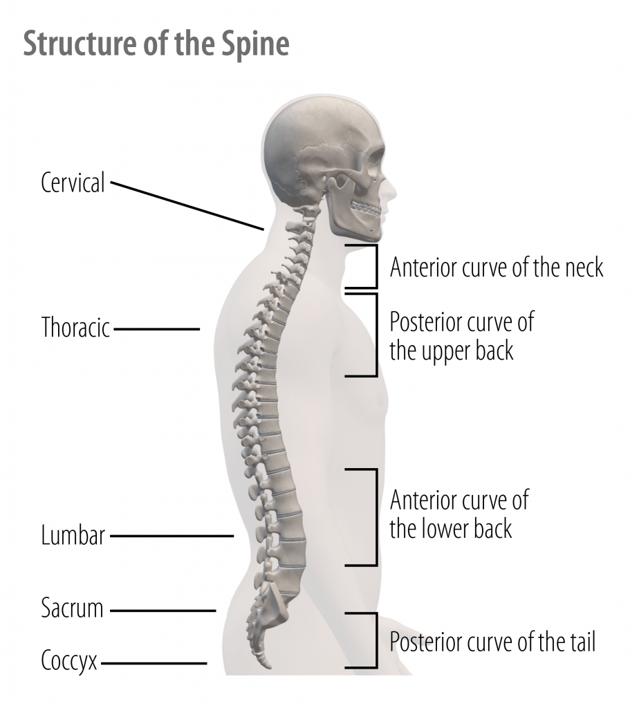
There are four natural curves of the spine: a kyphotic, or posterior curve of the upper back and tail, as well as a lordotic curve, or anterior curve of the lower lumbar spine and cervical spine.
There are 5 major regions of the spine:
- Cervical (C1-7) – The seven vertebrae in the neck form the cervical region of the spine. Cervical vertebrae are the thinnest and most delicate vertebrae in the spine but offer great flexibility to the neck.
- Thoracic (T1-12) – The 12 vertebrae in the chest region form the spine’s thoracic region. Thoracic vertebrae are larger and stronger than cervical vertebrae but are much less flexible. The spinous processes of the thoracic vertebrae point inferiorly to help lock the vertebrae together. A unique feature of the thoracic vertebrae is that each one forms joints with a pair of ribs to form the sturdy rib cage that protects the organs of the chest.
- Lumbar (L1-5) – The five vertebrae in the lower back form the lumbar region of the spine. Lumbar vertebrae are even larger and stronger than thoracic vertebrae but are more flexible due to the lack of ribs in the lumbar region. The lumbar spine carries most of our body- weight, which can lead to back problems in this region, despite the size and strength of the vertebrae.
- Sacral – The sacral region of the spine contains only the sacrum, a single bone in the adult skeleton that is formed by the fusion of five smaller vertebrae during adolescence. The sacrum is a flat, triangular bone found in the lower back and wedged between the two hip bones.
- Coccygeal – The spine’s coccygeal region contains only the coccyx, a single bone in the adult skeleton that is formed by the fusion of four tiny vertebrae during adolescence. The coccyx is often referred to as the human tailbone, as this region is homologous to the tailbones of animals that have tails. In humans, the coccyx bears our body weight when sitting down and provides attachment points for muscles of the pelvic and gluteal regions.
Joints are the point of articulation between bones. There are three main classifications of joints:
- Immovable (synarthrotic),
- Slightly moveable (amphiarthrotic), and
- Moveable (synovial, diarthrotic).
Joint composition varies by joint type. For moveable joints, the ends of the articulating bones are covered by cartilage. The articulation point is lined with a synovial membrane which reduces friction during movement. Ligaments are connective tissues that connect bones to bones and provide stability. Tendons attach the muscles to the bones.
While joints can be studied separately, they can be viewed as a series of joined parts that work together during movement. The term used to describe the parts of the body that work together, either in isolation or as a component of a more complex system, is kinetic chain. More complex chains of movements utilize multiple body parts at the same time. For example, in a squat, action occurs at the hip, knee, and ankle, while in a biceps curl, movement occurs at the elbow.
Instructor Tip: When working with older adults, instructors must recognize common skeletal system limitations and how these limitations affect movement demonstration. While encouraging safe movement demonstration, be prepared to accept a wide range of postural variations and movement ranges. Limitations in the shoulders and hip may produce irregular movement patterns while other conditions and limitations may limit a participant’s ability to use resistance equipment. Help each individual member achieve their goals.
Let’s examine some of the primary joints involved with movement.
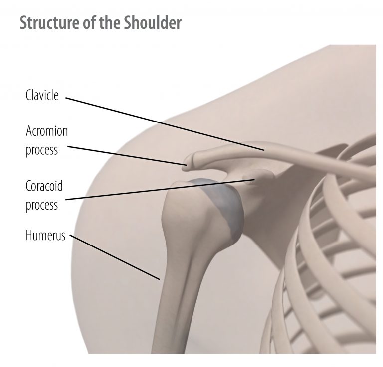
Shoulder Joint – The shoulder is a ball and socket joint, allowing movement in all directions. Actions include flexion, extension, adduction, abduction, rotation, circumduction, elevation and depression. The glenohumeral joint is the ball-and-socket junction of the top of the arm bone and the socket of the shoulder blade. The shoulder is versatile, but one of the least stable in the body. It depends heavily on ligaments for stability. The shoulders are involved in most upper-body exercises.
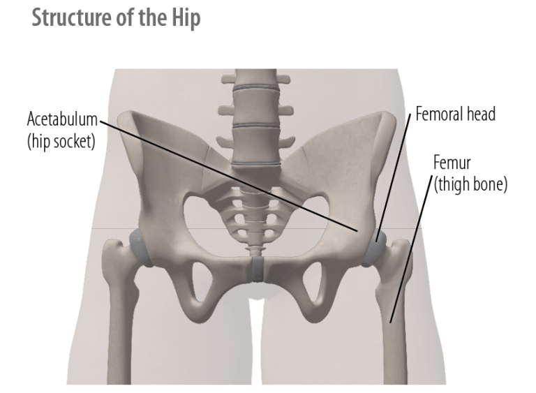
Hip Joint – The hip is also a ball and socket joint. The actions include flexion, extension, abduction, adduction and rotation. It’s large, stable and well supported. There are three main ligaments linking the femur to the hip joint. The hip is a kinetic chain from the back on one end and the knee, to the ankle and foot on the other end. The hips are involved in most lower body exercises and are a critical part of the gait cycle.
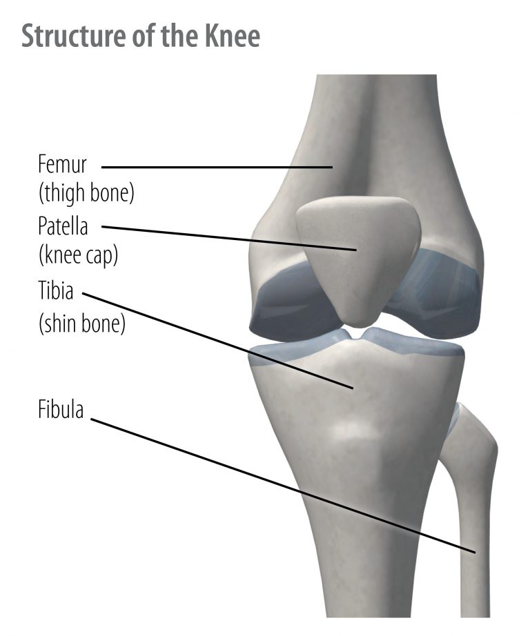
Knee – Hinge joints allow for movement in one plane. The knee is a large joint which includes three joint articulations: hinge, gliding (patella) and condylar (slight rotation). The main movements of the knee joint are flexion and extension. The ligaments of the knee provide stability and support. The anterior/posterior cruciate ligaments (ACL) prevent excess rotation, the medial/lateral collateral ligaments (MCL) prevent side bending stress, while the menisci cartilage serves as medial/lateral shock absorbers. The knee is a kinetic chain from the pelvis, hip, and upper leg on one end to the lower leg, ankle and foot on the other end.
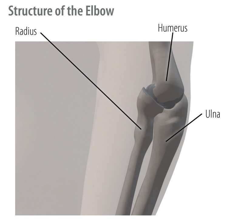
Elbow Joint – The elbow, like the knee, is a hinge joint. The elbow is also considered a pivot joint, which allows for rotation around a long bone. It flexes, extends and rotates over itself.
Thumb – The thumb is a saddle joint which permits movement in all directions. The human thumb is opposable, allowing the thumb to touch all fingers and grasp objects.
Ankle – The ankle is a saddle joint, allowing a simultaneous combination of movement and gliding in the tarsal bones. The ankle is supported by both ligaments and tendons.
The Muscular System
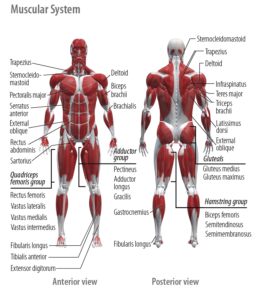
The body has three types of muscles:
- Cardiac – Heart muscle, contract automatically.
- Smooth – Involuntary muscles of the arteries which constrict and dilate to regulate blood flow, and intestines which require peristalsis to move food through the digestive tract.
- Skeletal – Voluntary muscles used for movement.
Skeletal muscle is composed of a multitude of muscle fibers. Connective tissue surrounds each muscle fiber, each fiber bundle, and the whole muscle. Connective tissue forms the tendon that attaches the muscle to the bone. A muscle fiber does not shorten or lengthen, as the muscle fiber runs the entire length of the muscle. Instead, muscles contract, similar to the way a rubber band lengthens or shortens.
Muscles contract when stimulated by a motor nerve, which is part of a motor unit. A motor unit consists of a single motor nerve and the individual muscle fibers that are activated by the nerve. When a motor nerve sends sufficient stimulus, the attached muscle fibers contract; the all or nothing principle. The number of motor units activated is dependent upon the amount of force that needs to be generated. Muscles produce the force that causes bones to move by attaching to at least two bones and crossing one or more joints. Muscles work together in groups to perform movement.
Instructor Tip: With age, your participants may be experiencing decreases in muscle mass, neuro-muscular control, and endurance. Be adaptable by providing multiple options for every exercise you teach.
Skeletal muscle allows for movement. There are two types of muscle fibers, slow twitch, and fast twitch. Individuals have a mix of slow and fast twitch fibers and muscle groups have varying ratios of fiber types. For example, the abdominal muscles continuously stabilize the torso and require low force over a long duration, thus, there is a higher percentage of slow twitch fibers.
Muscles work together in groups or pairs to produce movement and stabilization. Muscle groups may be superficial, or toward the surface, or deep within the body.
Upper Leg – The upper leg is made up of the anterior and posterior thigh muscles. Quadriceps, hamstring, and muscles of the hip, such as the hip flexors, and hip extensors are found in this region.
Lower Leg and Ankle – The lower leg is comprised of muscles that act on the knee and ankle joints. Major muscle groups include the anterior tibialis or shin, and the calf muscles.
Shoulder – The shoulder joint attaches the arm to the axial skeleton. The shoulder girdle is a complex group of joints and muscles supported by the muscles of the rotator cuff. These muscles are deep in the body and support movement of the shoulder and scapula. The shoulder’s superficial muscles produce movement of the upper arm.
Arm and Wrist – The arm attaches to the axial skeleton to produce upper body movement. The upper arm consists of large muscles of the biceps and triceps. The lower arm produces movement at the wrist.
Core – The abdominals and low back make up what is referred to as the core. The abdominal group consists of the rectus abdominis, internal and external obliques, quadratus lumborum and the transverse abdominis. Together with the back extensors, these muscles support posture and virtually every other movement of the body in a standing position.
Back – Muscles of the back play a key role in movement stabilization and activities of daily living. These muscles are comprised of the large muscles of the trapezius and latissimus, as well as smaller muscles that work to extend the arm.